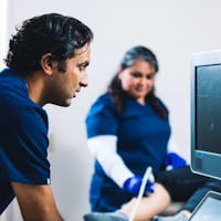
Ultrasound tests use sound waves to create images that help evaluate the heart, arteries, veins, or other areas of the body. At San Diego Cardiac Center, we offer several types of ultrasound tests in San Diego to support accurate diagnosis and care.
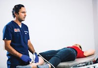
Carotid Ultrasound Ultrasound Test
The carotid arteries are on each side of your neck. These arteries deliver blood from the heart to the brain. A carotid ultrasound is performed to identify blocked or narrowed carotid arteries, which may pose a risk of stroke. It is a safe and pain-free test that involves putting a special gel on the area and moving the ultrasound device over your skin, which emits sound energy that creates images of the arteries and the blood moving through them. These arteries may be narrowed due to the buildup of plaque.